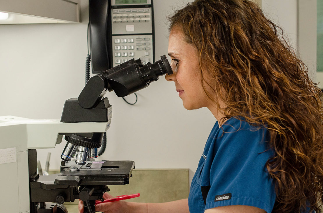
Using their knowledge and training, our dermatology providers diagnose and treat the majority of skin conditions by visual examination alone. However, in certain cases, additional information is helpful to differentiate between similar-appearing processes to make a more specific diagnosis (differentiating skin cancer from non-skin cancer or psoriasis from eczema, for example), or in order to guide more effective treatment. In these situations, a biopsy may be recommended for examination in our on-site laboratory by our in-house Board-Certified Dermatopathologist, Dr. Sara West or sent to local lab such as Regional Pathology Services.

What is a biopsy?
A biopsy is the process of removing a small sample from the affected skin. This may be done in one of three ways:
Shave biopsy – a small section of the skin is “scooped” from the surface and the area heals naturally.
Punch biopsy – a “cookie cutter” type of instrument is used to remove a small plug of skin and a suture or two are placed to close the wound.
Excisional biopsy – a scalpel is used to remove a slightly larger and deeper area of skin, generally in the case of a larger abnormal growth. Sutures are then placed to close the wound.
Regardless of the method, the area of skin to be removed will be numbed before the procedure. This ensures that there will be no discomfort during the removal. After the biopsy, the site will be covered with a topical antibiotic and a band-aid. Healing generally occurs within 2 – 3 weeks.
When is a biopsy recommended?
A biopsy is recommended when a visual examination alone is insufficiently precise to determine the nature of a skin condition. A biopsy may also be helpful to differentiate between two conditions that may appear very similar – for example, it can be used to distinguish between a cancerous skin growth and a non-cancerous skin growth or between psoriasis and eczema.
The decision to perform a biopsy is determined by the patient and provider together – it is important to weigh the expense of the procedure and the potential discoloration of the skin against the potential knowledge that can be gained from the testing.
Who examines the skin sample?
All biopsy samples are examined in DSO’s on-site laboratory by Dr. Sara West or sent to a local lab such as Regional Pathology Services. A Dermatopathologist is a physician who has extensive training in the microscopic examination of skin. The skin sample is embedded in wax, shaved into very thin slices, and mounted and stained on microscope slides. The dermatopathologist then examines the prepared slides to make a diagnosis that will guide selection of treatment options. The on-site laboratory allows DSO to have excellent quality control over the biopsy samples and microscope slides. This also ensures a shorter time from biopsy to diagnosis and treatment, since samples are prepared and examined without ever leaving DSO.
An amazing amount of information, unavailable in any other way, can be obtained by microscopic examination of a skin sample.
